Written by Areej Shahbaz
As molecular biology tools have gotten more affordable, dependable, and user-friendly, they are being used more frequently in laboratories. Since agarose gel electrophoresis is the most widely used technology for DNA molecule separation in various laboratories, its adoption as a research tool has been quick and widespread. This is due to the technique's simplicity and ease of understanding.
In this article, you will learn about the need for UV transilluminators with their innovation, importance, and requirement in the laboratories.
Let's first explore what you need to know about transilluminators before venturing further into the subject.
UV transilluminator

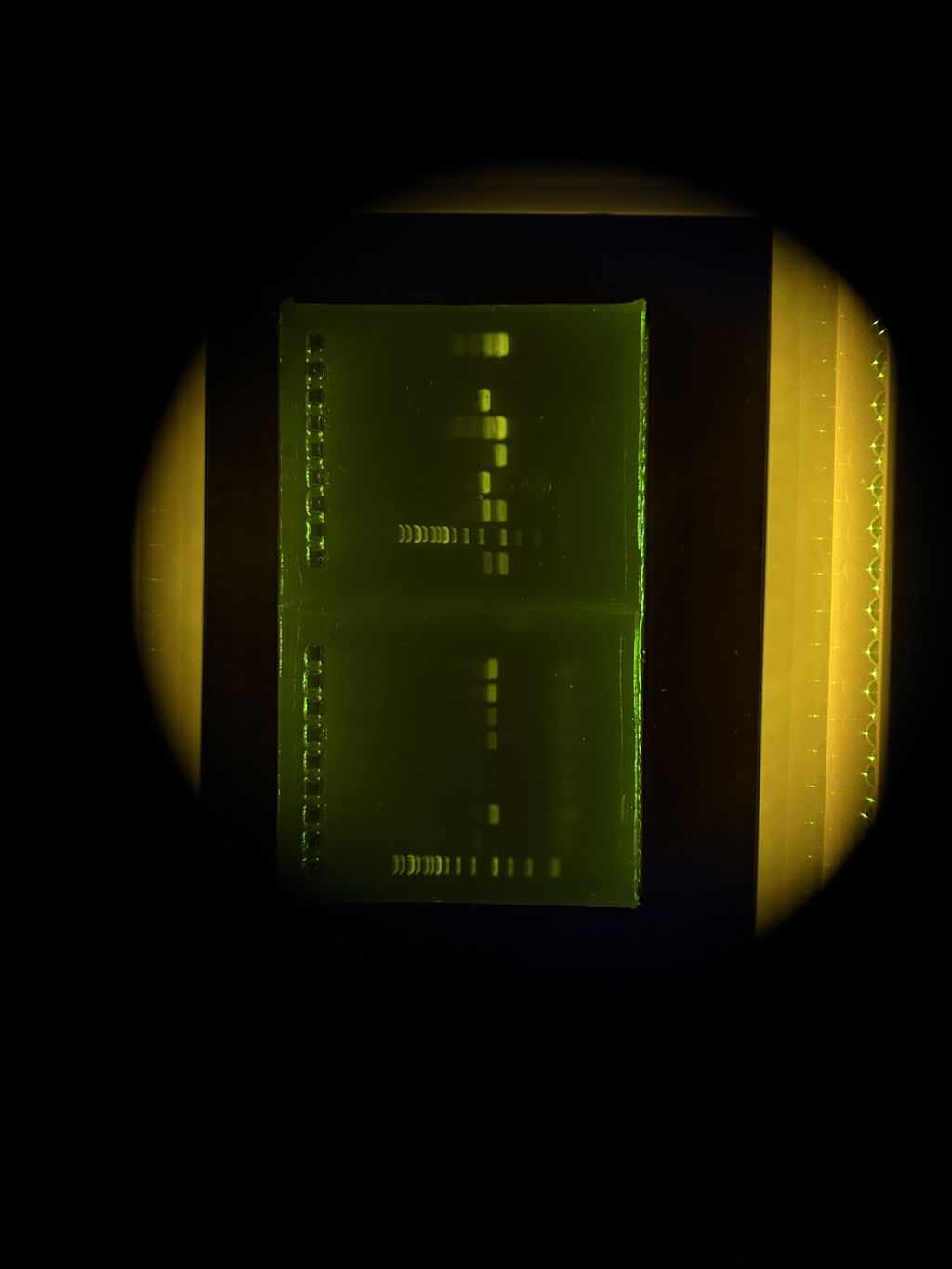
To see target oligonucleotides such as DNAs and peptides such as proteins in life science laboratories, an ultra-violet (UV) transilluminator is a common piece of equipment. To illuminate the wet agarose gels, UV transilluminators emit intense UV radiation through the viewing surface. A UV light source enables the fluorescent dye to fluoresce since the gels have already been stained with a fluorescent dye that binds to the nucleic acid.
The primary use of a UV transilluminator is for the post-electrophoresis viewing of DNA and protein agarose and polyacrylamide gels. Gels can be applied directly on the UV transilluminator; the wavelength will change depending on your specific application.
How do you select UV transilluminators?
When looking around for a UV transilluminator, some factors to bear in mind are viewing surface size for different size gels, wavelengths (as some models come in single, dual, or triple wavelengths), and the tabletop unit's dimensions.
UltraBright Blue/Green transilluminator - The Latest Innovation to LED Transilluminators
The most recent advancement in LED transilluminators is the UltraBright Blue/Green transilluminator. It is a top-of-the-line analytical system with cutting-edge construction for better performance and efficiency. With the UltraBright Blue/Green Transilluminator, it is possible to visualize bands on DNA or RNA gels stained with ethidium bromide, Gel green, SYBR Gold, SYBR Safe, SYBR Green I and II, Ruby, SRPRO Orange, SYPRO Ruby, Protein Gel stain, Coomassie Fluor Orange Stains, Green Fluorescent protein (GFP), Enhanced Green Fluorescence protein (EGFP), Lumitein and proprietary MaestroSafe pre-stain or loading dye for the UltraBright Blue/Green Transilluminator, which delivers strong excitation and low background.
Safer Blue/Green LED and simpler gel cutting beneath an amber filter are provided by the transilluminator. Higher sensitivity makes the majority of stains compatible. A wide range of light spectrums is used by UltraBright Blue/Green. (Blue) 475 nm and 520 nm (Green). The peak wavelength is therefore 505nm. Because of this, the UltraBright Blue/Green system is a particularly flexible one for the research lab.
Viewing of gels up to 200 mm by 160 mm is possible. The employment of a dichroic filter creates the best viewing conditions for band cutting and picture capture by considerably reducing background noise and enhancing band contrast.
Key Features
- Durable and elegant metal frame design.
- Simple to observe and cut gel.
- Safe to use i.e., blue LEDs don't cause skin or eye burns, nor do they cause UV-induced DNA damage.
- Light consistency is 5% which is most suited for visual analysis.
- It is convenient to use i.e., easy viewing of bands with a small design and a hinged changing position viewing screen. There is no need to wear amber glasses.
- High contrast and low background.
- Sensitivity down to 0.5-1 ng, designed for use with the majority of nucleic acid and protein stains.
- Intensity can be changed from 100% to 50%.
Specifications
- Light Source: Blue/Green LED
- Emission Wavelength: 505nm
- Design: Aluminum alloy metal frame with an amber filter for the blocking cover
- LED lifetime: approx. 50,000 hours
- Sample viewing area: (W × D) 200mm × 160mm
- Dimensions: (W × D × H) 340mm × 280mm × 80mm
- Power consumption: 24 W
- Electrical specifications: 100 ˗ 240 VAC: 50/60 Hz
- Sensitivity: down to 0.5-1 ng nucleic acid
- Light consistency: approx. 5%
- Weight: 3.8kg
Advantages
- There is no short wavelength radiation, so the user is safe.
- Blue and red dyes are exciting.
- All DNA stains are compatible.
- There is no DNA degradation.
Which dye is preferable to use and why?
Ethidium bromide (EtBr) and SYBR Green I are two nucleic acid gel stains that are frequently used in conjunction with UV illumination. SYBR Green I is a very weak mutagen that induces frameshift mutations, whereas ethidium bromide preferentially induces them in the presence of an exogenous metabolic activation system.
There are, however, safer alternatives to ethidium bromide. Green fluorescent cyanine dyes, such as SYBR Green, Accuris SmartGlowTM, and Apex Safe DNA Gel Stain, are highly sensitive and non-carcinogenic, according to the Ames test.
A highly sensitive stain for observing DNA in agarose or acrylamide gels is MaestroSafe or Invitrogen SYBR Green. SYBR Green stain is designed to be a less dangerous substitute for ethidium bromide and can be used in conjunction with either blue light or UV excitation.
Is SYBR Green the same as SYBR Safe?
Although the chemical makeup of each Invitrogen SYBR dye varies, they all share similar spectral characteristics. As a safer substitute for ethidium bromide, SYBR Safe DNA Gel Stain was created.
Blue/Green transilluminator is a superior technology
UV-light transilluminators visualize DNA by using a single wavelength. Red and green DNA dyes, such as ethidium bromide or Midori Green dyes, absorb well in the UV spectrum. This produces DNA bands of sufficient intensity; however, UV light is hazardous to both the user and the sample DNA. Just 30 seconds of UV-light exposure reduces cloning efficiency significantly, which has implications for downstream applications. As a result, UV-light visualization of DNA is not the preferred method. In contrast to UV light, Blue/Green LED technology employs a broad spectrum of light ranging from 470 nm to 520 nm. This light does not harm DNA or the user. Even ethidium bromide or other red DNA dyes with low spectral absorption show DNA band intensity comparable to UV illumination.

This is due to the DNA's accumulated energy absorption (area under the curve) in the Blue/Green spectrum. Green DNA dyes have a very high absorption intensity in the Blue/Green light spectrum, resulting in more intense DNA bands.
Illuminators with blue and green LEDs aren't simply for gels. Almost everything that fluoresces green or red can be detected and imaged in your lab. You can triple the effectiveness of your subcloning transformations by using safe blue/green LED illumination.
For a complete guide, see our Definitive Guide to Blue Light Transilluminators